
TBI and Degenerative Brain diseases: What are they?
Hi everyone! So just a bit of a heads up, the format we’re going to be following in the next several articles is going to be a bit different. Each article is going to be a about a specific aspect of health and will eventually all culminate in how all these aspects relate to a specific medical condition. It’s important to me that these articles all be stand-alone for future reference, so the articles will be fairly broad to start off on, with definitions for many terms as footnotes. If you have further questions, you can always comment, OR Google is your friend. That being said, let’s begin! This first part in the series is about what are the commonalities about Traumatic Brain Injuries (TBI) and Degenerative Brian Diseases (DBD) and what’s happening to your brain while this is going on.
These days, the more violent the sport, the more popular it’s becoming. Activities like MMA, UFC, kickboxing, and football are everywhere you look. But there’s something else in common with all of these activities – TBI. There’s currently a plethora of studies and attention being brought to this issue due to the release of movies like “Concussion”, and the general attention brought to the danger of football. Here’s the catch – a lot of the symptoms of TBI mirror those of DBDs, like Alzheimer’s and Parkinson’s.
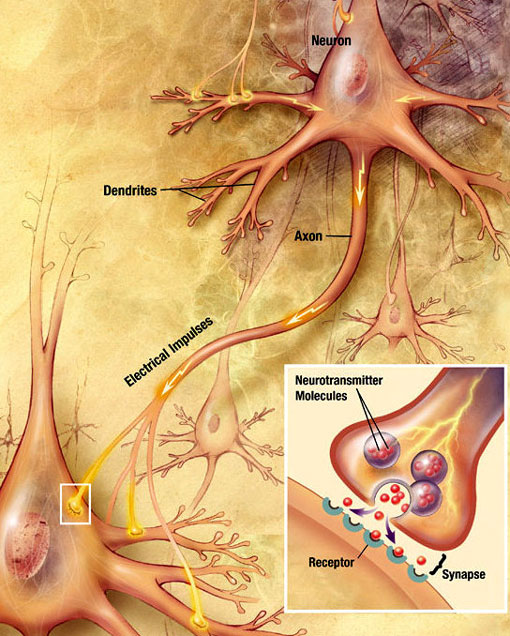
So what exactly happens with a TBI? When you get hit in the head too much, or really at all, the brain takes on mechanical damage, injuring its tissues, and also increases blood flow to the area. This leads to a chain reaction of feedback in the brain. The first is the accumulation of lactic acid due to anaerobic glycolysis¹. The ATP stores become depleted and this leads to a breakdown of the ion pumps in the brain. Simultaneously, increased membrane permeability, or the damaging the protective tissues around the brain is taking place. This causes the blood-brain barrier to become “leaky”, which causes molecules like potassium and chlorine as well as neurotransmitters like glutamate and aspartate to flood the brain. At the same time, the calcium-sodium pumps, which became dysfunctional during the first steps, become imbalanced and the combination of all of these reactions leads to a depolarization in the system, which causes swelling in the brain. Simultaneously, the immune system in the body kicks in when the tissue in the brain is damaged, and micro-gleal¹ cells are activated and migrate to “plug up the holes” in the brain membrane. This is what causes the swelling in the brain. This swelling, can eventually lead to an edema² – the third step. When an edema forms, the extra calcium ions lead to an increased concentration of free radicals and fatty acids. The structure of the biological membranes and the nucleosomal DNA also changes. During this same process, the micro-gleal cells are trying to eat cellular debris that occurred during the damage and this causes neural-inflammation. These three steps together lead to the degradation of the membrane and ultimately, programmed cell death, a reaction the body has to destroy potentially pre-cancerous cells. All of this happens within a few hours of the initial injury. What we end up with as the final product is the accumulation of Amaloyd-Beta Plaques (ABPs). (1 and 2)

fighter with cerebral edema

Normal Brain vs. Alzheimer’s Brain PET Scan
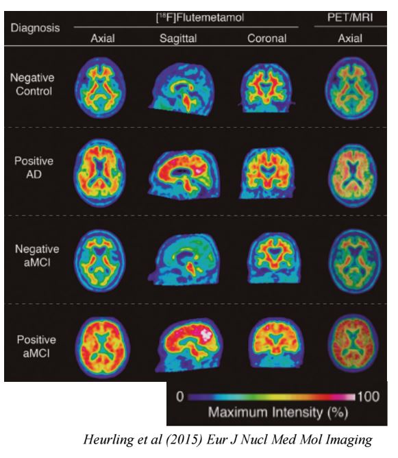
Normal Brain vs. Alzheimer’s Brains

Process by which dementia takes place
And finally, what is Parkinson’s? Parkinson’s is a progressive neurodegenerative disease, which inhibits overall movement, due to the death of nerve cells. Parkinson’s mainly affects dopamine in cells in the part of the brain called the “substantia nigra”³, whose dying neurons produce less and less dopamine as the disease progresses. Furthermore, the presence of alpha-synuclein proteins, or Lewy bodies, in the mid-brain, brain stem and olfactory bulb are also indicative of Parkinson’s. There are 4 main indicators of Parkinson’s: tremor of the hands, arms, legs, jaw and face, bradykinesia or slowness of movement, rigidity or stiffness of the limbs and trunk, and postural instability or impaired balance and coordination. (5) There, however, is another type of Parkinson’s known as Parkinsonism. It is a disease that exhibit the 4 symptoms of Parkinson’s, but is not Parkinson’s. According to www.atrainceu.com,
“Parkinsonism often has an identifiable cause, such as exposure to toxins, methamphetamine, trauma, multiple strokes, other nervous system disorders, or illness. Generally, Lewy bodies are not seen in parkinsonism. The term parkinsonism is also associated with disorders such as progressive supranuclear palsy, multiple system atrophy, Lewy body dementia, corticobasal degeneration, vascular parkinsonism, drug-induced parkinsonism, and parkinsonism secondary to infection and other causes (Hohler et al., 2012)”
Interestingly, methamphetamine users often have Parkinsonism and are at a higher likelihood to develop Parkinson’s. Now, the exact pathopsychology of Parkinson’s is not completely known. What we do know is that several factors cause Parkinson’s. First off is the most commonly known reason – the lack of dopamine. Dopamine is secreted into the synapse, crosses the synapse, and activates dopamine receptors. Unused dopamine is absorbed back into the presynaptic membrane, to be released next time. However, as an individual ages, they produce less and less dopamine. This lack of dopamine results in cell death. However, in Parkinson’s this process is accelerated. The decrease of dopamine also causes an eventual decrease in the production of glutamate, GABA, and serotonin. Dopamine decrease is not the only indicator of Parkinson’s however. As aforementioned, Lewy bodies are also present in Parkinson’s patients. Lewy bodies are an cluster of misfolded proteins that abnormally form within the neuron. How these proteins are related to Parkinson’s however, is uncertain. (6) As for Parkinsonism, the pathopsychology is unclear.
How dopamine travels through the neuron

Dopamine levels in a normal person vs. Parkinson’s patient

Dopamine levels in normal individuals vs. Parkinson’s patients

PET Scan of Normal Brain vs. Parkinson’s Brain
So what do all these disease have in common? If you’ll notice, all of the diseases end up with the production of ABPs. According to http://www.atrainceu.com
“Neurodegenerative disorders such as Alzheimer’s disease, frontal-temporal degeneration, prion disease, Huntington’s chorea, and motoneuron diseases are increasingly being realized to have common cellular and molecular mechanisms, including protein aggregation and inclusion body formation in certain areas of the nervous system.”
Essentially, the breakdown comes to this: When the brain experiences any kind of “stress”, whether that be external or internal, it begins to create ABPs. The creation of ABPs leads to a negative feedback cycle of more ABPs being created, and eventually, cell death in the brain. When brain cells are stressed, they release a protein that tells them to die. Sometimes, they don’t die immediately, but the more times they are stressed over time, the more “death targeting” proteins they release. This makes the cells more susceptible to death, eventually killing off large groups of cells that have undergone repeated stress. This process predisposes individuals to neurodegenerative diseases. TBIs and DBDs all have this in common: a large amount of ABPs and Tao proteins coming together to inflame parts of the brain. The only reason many exclude Alzheimer’s from the neurodegenerative diseases is because it is specifically age related, though as previously discussed, TBIs can affect the time-span in which you get a DBD. (1) Long story short, there’s still a lot to learn about DBDs and TBIs, but inflammation is generally bad. Be ready to learn more in the next article!
Footnotes:
- Anaerobic respiration is the process of producing cellular energy without oxygen. Anaerobic respiration is a relatively fast reaction and produces 2 ATP, which is far fewer than aerobic respiration.

Anaerobic and Aerobic respiration
- Microglia are a type of glial cell that are the resident macrophages of the brain and spinal cord, and thus act as the first and main form of active immune defense in the central nervous system (CNS). Microglia constitute 10–15% of all cells found within the brain
- An Edema is a condition characterized by an excess of watery fluid collecting in the cavities or tissues of the body.
- Substantia nigra is a brain structure located in the mesencephalon (midbrain) that plays an important role in reward, addiction, and movement.

Substantia nigra in Normal brain vs. Parkinson’s brain
Sources:
- Joe Rogan interview with Rhonda Patrick: http://podcasts.joerogan.net/podcasts/dr-rhonda-patrick-3
- http://bja.oxfordjournals.org/content/99/1/4.full
- Lectures by Enrique Cadenas
- Science Friday: http://www.sciencefriday.com/segments/always-hungry-your-fat-cells-may-be-to-blame/
- http://www.pdf.org/about_pd
- https://www.atrainceu.com/course-module/2441043-143_parkinsons-module-02
- http://www.traumaticbraininjury.com
- http://www.mayoclinic.org/diseases-conditions/traumatic-brain-injury/basics/symptoms/con-20029302
- http://www.cdc.gov/traumaticbraininjury/get_the_facts.html
- http://www.apdaparkinson.org/parkinsons-disease/understanding-the-basics/
- http://www.clevelandclinicmeded.com/medicalpubs/diseasemanagement/neurology/parkinsons-disease/
- http://www.ncbi.nlm.nih.gov/pmc/articles/PMC2467461/
Elephant Seals – Worse Than Your Akita’s Shedding
As aforementioned, I’ve currently become deeply invested in podcasts. One such podcast is “Science Friday” hosted by Ira Flatow on Public Radio International. On a recent jam-packed episode, an exciting topic came up – Elephant Seals [1]! These (not-so) little guys have been causing lots of problems for the last couple of decades; and we’re only finding out now!
First, a little bit about Elephant Seals. There are two breeds of Elephant Seals – Northern and Southern [2]. In this study, we focused on the Northern breed or Mirounga angustirostris. The Southern species is not only significantly larger, but also lives longer than its Northern counterpart. The Northern species lives only along the Pacific coast of Northern America. In terms of IUCN status, the Mirounga angustirostris is a success story! Once thought to be extinct due to over-hunting, this guy has made a comeback and is back up at around 120,000+ [3].

In case anyone was wondering why they’re called “Elephant” Seals… (Mirounga angustirostris)

Mirounga leonine (Southern Elephant Seal)

Elephant Seal shedding

Elephant Seal puppy
For several decades, scientists have been noticing severe mercury spikes in the Pacific Ocean off the coast of California. The case was unknown for years until an exciting development came along in science – using DNA mapping technology to map the DNA of the ocean! What was the result? This mercury was coming from the coast? Lo and behold, we find shedding Elephant Seals on the shoreline!
During a recent study (which can be found here [4]) at UC Santa Cruz, it was discovered that during the molting season, levels of methyl-mercury – one of the most toxic forms of mercury – was 17 times higher than normal (as if it wasn’t dangerous enough in the body already). And it’s in fact precisely because of the mercury’s poisonous levels that Elephant Seals try to shed their hair. The Seals will cumulate the mercury in the hair on their bodies, and then shed the hair to rid the body of the toxin [5], in a process called Catastrophic Molting [6]!
One might ask how we know that the mercury is coming from the Elephant Seals fur and not from their feces or other fauna altogether. The answer lies in newborn Seals. Because mercury is so easily absorbed into the system of living creatures, it stands to reason that pregnant mothers would “infect” their unborn children as well. In fact, pups born to contaminated mothers showed high levels of methyl-mercury in their “natal coal” or the hair that they are born with [7]. So, we can extrapolate that the seals coat is to blame for increased ocean methyl-mercury levels.
Now you might not think that this is a problem, but the mercury in the hair gets washed up back into the oceans, digested by microbes in the water, and moves its way back up the food chain, until it may have serious consequences for humans, Elephant Seals, and the ecosystem overall. The levels found around Seal molting grounds are higher than those found in highly urbanized, contaminated coastal towns. This problem is further accentuated by the fact that industrial pollutants in the water have already significantly increased mercury levels in the oceans.
What makes the situation worse is that mercury doesn’t degrade either, which means that overtime, it’s going to concentrate itself more heavily at the top of the food chain, with unknown consequences. This process is known as biomagnification.
So, do we know anything? Well, yes. We know the harmful effects that methyl-mercury has on the human body. According to the EPA, methyl-mercury has significant effects on humans, including but not limited to: neurological development in infants, “impairment of the peripheral vision; disturbances in sensations (“pins and needles” feelings, usually in the hands, feet, and around the mouth); lack of coordination of movements; impairment of speech, hearing, walking; and muscle weakness” [8] and even potentially death. One such extreme case of mercury poisoning was in Japan, from 1932 to 1968 [9]. A factory that produced acetic acid discharged its waste into Minamata Bay, where nearby residents consumed contaminated shellfish for years. What was eventually known as Minamata disease caused “brain damage, paralysis, incoherent speech and delirium” in over 50,000 local residents.
So, how do we stop all of this? Scientists aren’t quite sure, but one thing is for sure: we need to reduce our pollutant footprint if we want to keep these cute guys around, and other apex predators; especially if we want a chance of surviving a healthy human life, for us and generations to come.
Sources:
- http://www.sciencefriday.com/segment/09/11/2015/testing-ocean-dna-americans-pass-a-science-quiz-and-polar-bear-diets.html
- http://animals.nationalgeographic.com/animals/mammals/elephant-seal/
- http://www.nmfs.noaa.gov/pr/species/mammals/pinnipeds/northernelephantseal.htm
- http://www.pnas.org/content/early/2015/09/02/1506520112
- http://www.santacruzsentinel.com/environment-and-nature/20150917/molting-elephant-seals-recycle-mercury-back-into-seawater
- http://www.mnn.com/earth-matters/wilderness-resources/stories/elephant-seals-are-raising-mercury-levels-california-beach
- http://www.theverge.com/2015/9/12/9313363/mercury-molting-seals-fur-California
- http://www.epa.gov/mercury/effects.htm#meth
- http://www.who.int/mediacentre/factsheets/fs361/en/
